MEA Recordings

Next Generation Cellular and Organotypic Assays
Principal Senior Scientist
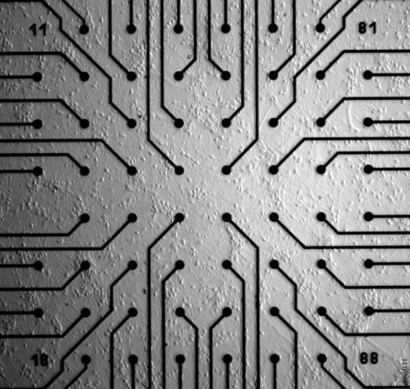
MEA (microelectrode array) offers extracellular recording with optional optical or electrical stimulation. It is performed on electrogenic tissue on the top of substrate integrated electrodes.
MEA shows certain benefits over other methods in electrophysiology:
- Information on the network level
- Non-invasive
- Long term recordings possible with cell culture possible for days or weeks
- “holistic” ion channel interactions
Our Services
- safety pharmacology (seizure effects on neurons, arrhythmia on cardiac cells)
- neurotoxicity; neuronal modulation; LTP (long term potentiation)
- neurodegeneration (organotypic cultivation on brain slices/retina)
- compound MoA
- diabetes research
- retina research
Methods
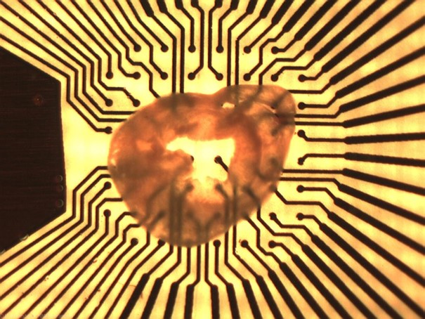
MEA recordings can be performed using different models such as neurons, cardiomyocytes, brain/cardiac slices, iPSC-derived neurons/cardiomyocytes etc.
Instruments
Different MEA systems are available depending on the application (high number of electrodes e.g. for neuronal applications, high-throughput systems (96-well format), 96-channel pipettor for parallelized substance application).
NMI Natural and Medical Sciences Institute
at the University of Tübingen
Markwiesenstraße 55, 72770 Reutlingen
Tel.: +49 7121 51530-0
Mail:
info@nmi.de
Follow us now