Microtumor Models

Group Leader Tumor Biology
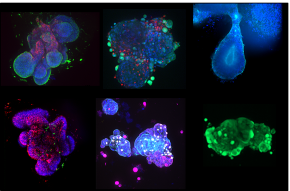
Patient-derived microtumors for drug development and compound efficacy testing
By developing novel patient-derived cellular model systems, our Tumor Biology Group supports preclinical efficacy testing and validation of pharmacological agents, as well as biomarker analysis for diagnostics and therapeutic outcome monitoring, in collaboration with academic and clinical researchers, as well as customers and partners from industry.
Our services
Test systems for cancer biology:
- Ex vivo microtumor models (established for different tumor types)
- Primary immune cells (T lymphocytes, TILs, PBMCs)
- Microtumor-immune cell co-cultures
- Variable culture formats (e.g. 96-well)
Methods
- Immunohistochemistry analysis of tumor tissue and ex vivo microtumors (H&E, automated DAB based target protein/marker staining).
- Wide-field and confocal microscopy, slide-scanning microscopy, fluorescence microscopy
- Live cell imaging and image-based analysis
- Functional assays: including cytotoxicity, proliferation, immune cell killing, apoptosis
- FACS analysis: including multiplexed immune cell phenotyping
- Bead-based multiplexed immunoassays for analysis of secreted immune mediators (including chemokines/cytokines)
- Gene expression analysis by qRT-PCR
Available technologies
- HistoCore PEARL - Tissue Processor
- Dako Autostainer Link 48 with pretreatment module
- ZEISS Axioscan Microscope Slide Scanner
- Zeiss Cell Observer Spinning Disk Confocal Microscope
- BD LSRFortessa Cell Analyzer Flow Cytometer
- Multimode microplate reader for fluorescence and luminescence-based assays
- Fully equipped histology laboratories as well as cell culture laboratories of safety level 1 and 2
NMI Natural and Medical Sciences Institute
at the University of Tübingen
Markwiesenstraße 55, 72770 Reutlingen
Tel.: +49 7121 51530-0
Mail: info@nmi.de
Follow us now